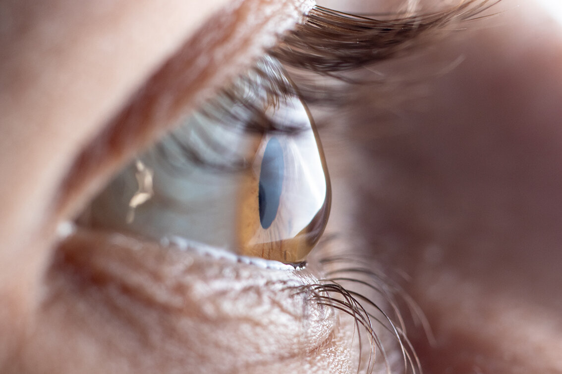
Keratoconus
What is Keratoconus?
The cornea is the clear window at the front of the eye that is essential for good vision as it helps to focus light. The shape of the cornea is important and being round like a football allows the cornea to focus light perfectly. In keratoconus, the cornea gradually loses its strength and then changes shape. As it does so, your clear vision (focus) is lost. Most cases of keratoconus will gradually worsen until it stabilises around 40 years of age.
What are the symptoms of Keratoconus?
The effect of keratoconus on your vision depends on its severity. You may notice an increased sensitivity of light in the early stages. As the shape of your cornea changes, you may have to frequently change your glasses prescription to maintain good vision. There is a limit to the improvement that glasses will achieve and as the keratoconus progresses, your vision will become blurred despite wearing glasses.
What are the treatments for Keratoconus?
Treatment of keratoconus has 2 aims:
Shape stabilisation - To stop your cornea from continuing to lose its shape, and
Visual rehabilitation - To improve your vision.
There are some very simple things you can do to help prevent your keratoconus deteriorating. It has been shown that repeatedly rubbing your eyes may weaken the cornea; it is therefore vital to stop rubbing your eyes even after collagen crosslinking. This may be difficult if you suffer from itchy or irritated eyes. To assist it is important to treat any conditions such as allergic eye disease and hay fever that may be contributing to this. We are happy to review and help you manage this.
Corneal collagen cross-linking (CXL) aims to stabilise and strengthen the cornea and preventing the keratoconus from worsening. This treatment is very effective halting progression in >80-93% of corneas. The lower success rate is more commonly seen in younger patients and those with more advanced disease. CXL does not improve the quality of vision but may lead to a change in your contact lens or glasses prescription.
The method used to improve your vision depends personal preference and the stage of your keratoconus. Very mild cases may require glasses or soft contact lenses. As the shape of the cornea becomes more deformed, hard contact lenses may be required. Other surgical alternatives to this include customised laser eye surgery or the insertion of intrastromal rings to support the cornea. In advanced cases of keratoconus, a corneal graft may be required.
Mr. Darcy offers the entire range of treatments and will guide you through this process.
Why does keratoconus occur? +
The cornea has five layers. Keratoconus develops when there is a structural weakness in these layers. The reason for this is still not fully understood, but it is likely to be a combination of genetic (passed down in families) and environmental causes. For example, people with allergies are more prone to keratoconus, particularly with eye rubbing.
The natural ageing process of the cornea means that by around the age of 40 most people with keratoconus will not progress any further. Some other rarer causes of corneal warpage can still progress beyond the age of 40; Pellucid marginal degeneration and post-laser ectasia.
How do I know if I have keratoconus? +
Keratoconus typically starts in the early teenage years but can present in both younger children or adulthood. Keratoconus always affects both eyes, but the changes do not usually occur at the same speed (2). One eye can remain relatively normal, meaning deterioration of one may be unnoticed if the fellow eye remains good.
In the early stages, you may notice increased sensitivity to light, especially bright lights such as car headlights. To overcome this light sensitivity, children may start to squint or partially close the eyes. As keratoconus develops the blurring worsens. In children, this may become apparent with them having increasing difficulty seeing the front of the classroom.
Often the first thing people notice is they need to change their glasses with increasing frequency (changing more frequently than every two years).
Who is at risk of getting keratoconus? +
Overall, 55 of every 100,000 people have keratoconus, but this is higher in certain groups (3). It is more common in certain genetic conditions; Down's syndrome, Ehlers-Danlos, Marfans (4). There is also a link with other eye conditions; Fuch's Endothelial Dystrophy, Leber's congenital amaurosis, retinitis pigmentosa, and retinopathy of prematurity.
Keratoconus is more common in people with hayfever, asthma and other allergies. It is essential to treat any allergic eye disease. Allergic eye disease causes your eyes to become itchy and red. It is vitally important to stop any rubbing and itching your eyes, which, can worsen keratoconus. This can often be very difficult. Many anti-allergy drops are available over the counter, and your optometrist/GP may also suggest some. Oral anti-allergy medication can also be helpful. Some less sedating oral preparations are only available through a doctor. If these do not resolve your symptoms, then you will need specialist advice. Some medications are only available through a specialist ophthalmologist. We have years of experience and will advise you of the most appropriate treatment.
If you have keratoconus, it is vital to be assessed by a Corneal Specialist. We have many years of experience treating and managing keratoconus.
How is keratoconus diagnosed? +
Keratoconus can be very difficult to detect in the early stages. The blurring of the vision is often very gradual, and so people may not notice. Your optometrist may be the first to suspect it by detecting subtle signs while performing an eye test. Everybody must see an Optometrist at least every two years.
We use specialist equipment to detect keratoconus, which is very sensitive and can pick up even very early changes. The same tests are then used to monitor your keratoconus.
How often will I need to be seen for my keratoconus? +
This varies and depends upon; the stage of the disease, whether there are signs of progression, the persons' age. In general, stable patients need reviewing every 6-24 months, whereas those with signs of progression will require more frequent assessments.
Can I wear my contact lenses to the appointment? +
Contact lenses are very effective at improving the vision in many people with keratoconus. They often give a better quality of vision than glasses. Unfortunately, contact lenses temporarily change the shape of the cornea and can hide some of the changes seen in keratoconus. It is therefore essential to remove soft contact lenses one week before the appointment. Hard lenses (e.g. RGP) need to be removed four weeks before the appointment. We understand this may be very difficult or even disabling for some people. If so, please contact us, and we will assist in finding a suitable solution.
Can I switch to from hard to soft contact lenses before my appointment? +
This is often a good solution for many hard contact lens wearers. It is still crucial to have a week out of the soft lenses before your appointment.
Can I have my eyes assessed on different days so I can keep wearing a contact lens in one eye? +
We often find this is the most convenient way method people, particularly with advanced keratoconus. Additional fees will apply.
What are the surgical treatments for keratoconus? +
Except for collagen cross-linking, all of the surgical treatments are performed with the intent of improving the eyesight. There are usually several different options available. Mr Darcy offers a comprehensive range of treatments and will guide you through this process, providing a bespoke treatment plan.
What are kerarings? +
Kerarings are acrylic rings that are surgically implanted and used to re-support the cornea. Kerarings aim to improve the quality of your eyesight by improving the shape of the central section of the cornea that we predominately look through. Inserting a keraring is akin to replacing a broken tent pole. For more information (hyperlink to keraring page)
Can I have laser eye surgery? +
Keratoconus and other ectasias are contra-indications for traditional laser eye surgery, that is designed to remove the need for glasses or contact lenses. We use laser eye surgery in keratoconus, but the aim is to improve the quality of your vision in glasses or contact lenses and not to remove the need for them. The irregular shaped cornea caused by keratoconus causes scattering of light that results in symptoms such as starbursts, halos and glare. We use the laser to reduce these effects. Not everybody is suitable for this treatment. We will assess your eyes and advise whether you may benefit.
Will I need a corneal graft? +
The number of corneal grafts performed for keratoconus has decreased since the introduction of collagen cross-linking (5). They are, however, still an excellent option for people whose vision cannot be improved by alternative methods. If required, Mr Darcy will guide you and advise of the most appropriate type of graft.
Advanced Learning Zone
How is the progression of keratoconus defined? +
Worsening of keratoconus is called progression. As the cornea further warps, it becomes steeper in some areas. In these areas, the tissue is stretched, causing it to thin. The steepening is classically very irregular and usually starts on the inside surface of the cornea. Specialist machines are used to measure the shape of the inside and outside corneal surfaces. The relative thickness of each part of the cornea is also measured. Your glasses prescription, and vision are also considered, to complete the picture. Your eyesight is assessed objectively with a visual acuity chart, but we also consider your subjective assessment of your quality of vision.
How accurate are the machines used to measure keratoconus? +
There are two types of machines used; Pentacam and an OCT. We use the measurements from both because all devices have some 'noise' in their measurement accuracy. In keratoconus, the amount of noise increases with more advanced disease. To determine progression, we look for a change in the shape of the front or back surface, or the cornea becoming thinner (5). These changes must be outside of the 'normal noise' of the testing system.
How do contact lenses change the shape of the eye? +
The outside layer of the cornea is called the epithelium. It is a tough outer skin that is continuously reproduced to maintain corneal integrity. In the centre, it is usually ~50µm thick, similar to the thickness of a human hair, representing ~10% of the total corneal thickness. The epithelium has an inherent ability to alter its thickness, creating a smoothing effect, so that over peaks, it thins and depressions, it thickens. We use this in the early stages of keratoconus to aid diagnosis.
Contact lens wear also causes the epithelium to change in thickness. They usually have a smoothing effect, which partially flattens any peaks. Accurately measuring the peaks is essential in determining whether keratoconus is progressing. Contact lenses can, therefore, 'hide' some of the changes caused by keratoconus. After contact lenses removal, the effect on the epithelium slowly reverses, classically taking one week for soft lenses and four for hard lenses.
What is the difference between keratoconus, Pellucid Marginal Degeneration (PMD) and Keratoglobus? +
All three are ectasias. The features that distinguish them are the location of the thinning and the pattern. Keratoconus and PMD are considered variants of the same disease. Keratoglobus is considered its own entity.
References
Williams KM, Verhoeven VJM, Cumberland P, et al. Prevalence of refractive error in Europe: the European Eye Epidemiology (E(3)) Consortium. Eur J Epidemiol. 2015;30(4):305-315. doi:10.1007/s10654-015-0010-0
Gomes JAP, Tan D, Rapuano CJ, et al. Global Consensus on Keratoconus and Ectatic Diseases. Cornea. 2015;34(4). https://journals.lww.com/corneajrnl/Fulltext/2015/04000/Global_Consensus_on_Keratoconus_and_Ectatic.1.aspx.
Kennedy RH, Bourne WM, Dyer JA. A 48-year clinical and epidemiologic study of keratoconus. Am J Ophthalmol. 1986;101(3):267-273. doi:10.1016/0002-9394(86)90817-2
Cullen JF, Butler HG. DOWNS SYNDROME AND KERATOCONUS. Br J Ophthalmol. 1963;47(6):321 LP - 330. doi:10.1136/bjo.47.6.321
Godefrooij DA, Gans R, Imhof SM, Wisse RPL. Nationwide reduction in the number of corneal transplantations for keratoconus following the implementation of cross-linking. Acta Ophthalmol. 2016;94(7):675-678. doi:10.1111/aos.13095

