
Customised laser eye surgery
Any corneal irregularity degrades the quality of vision. Technological advances with phototherapeutic keratectomy laser eye surgery allow accurate reshaping, restoring vision.
Restoring vision with customised laser eye surgery
The cornea must be optically transparent and have a regular, smooth shape for clear vision. Any irregularity causes an increase in the blurring of vision through scattering light. There are many reasons why a cornea may not have an ideal shape, including;
- Injuries or trauma
- Corneal ectasia - keratoconus, Pellucid marginal degeneration
- Previous surgery
- Genetic conditions

Understanding imaging
We use differing imaging techniques to assess eyes and the quality of vision. Combined, these offer a powerful tool. Mr Darcy has carefully selected a combination of imaging techniques to provide comprehensive coverage at Vision Care Clinic. We often deliberately use different technologies to measure the same part of the eye. This provides invaluable second or even third checks of the information.
Corneal Tomography
We use highly accurate machines to measure the shape of the cornea. These produce contour maps similar to an Ordinance Survey map used for walking.
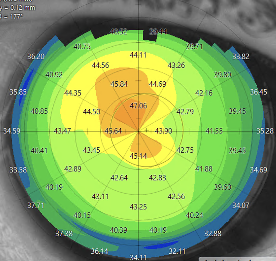
A regular shape should either be uniform in colour or have a figure of eight shapes, signifying astigmatism.

Normal cornea
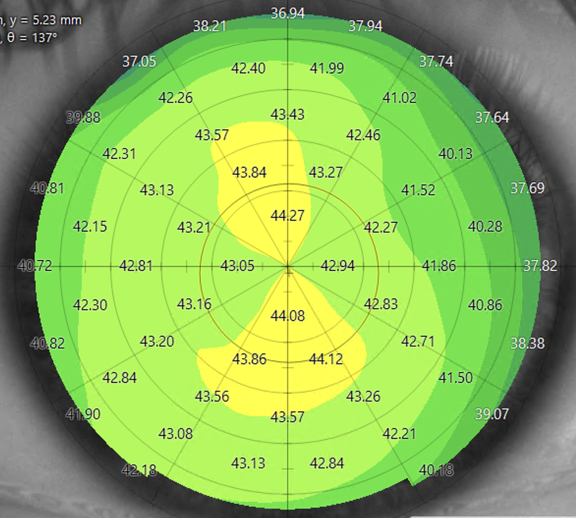
Normal cornea with regular astigmatism
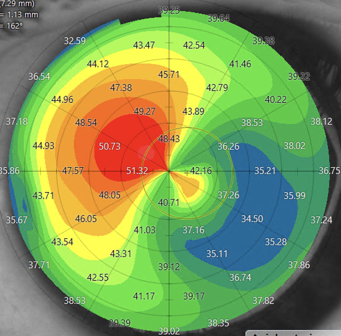
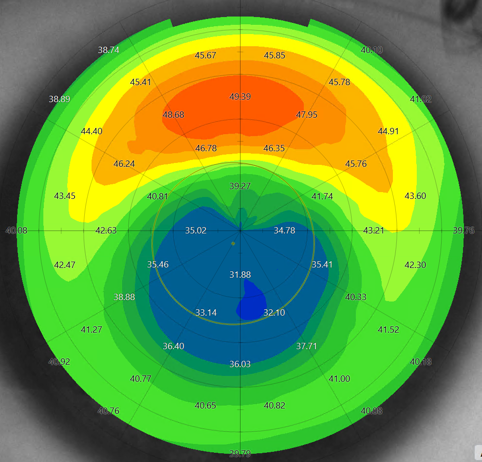
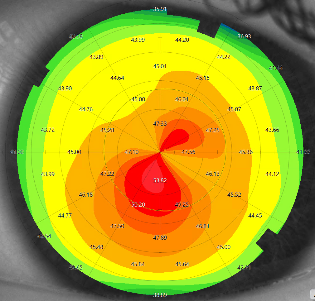
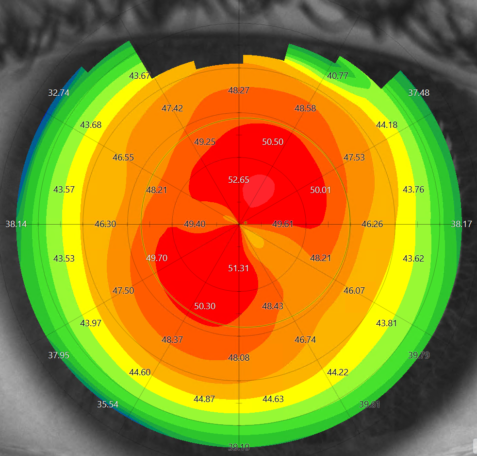
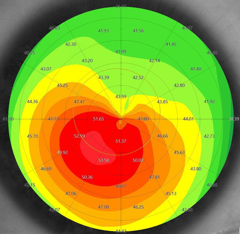
The colors in the scans indicate elevation. If we use a walking analogy, the green and yellow areas represent flat lands. The slope becomes steeper as it transitions from yellow to orange and then red. The steepest areas appear in a white colour, similar to snow-tipped mountains.
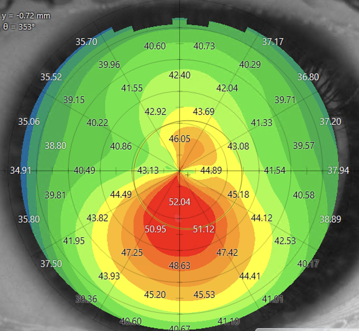
Keratoconus
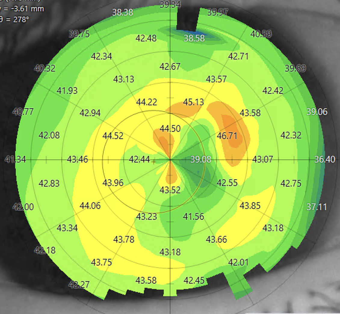
Corneal scar
Rapid color changes over small areas occur where there is significant irregularity. The yellow and black circle indicates the pupil. Irregularities in this central area affect vision more significantly.
Aberrometry
This specialist machine shines a ray of light into the back of the eye. The retina then reflects this ray, bouncing it back out of the eye. The machine can capture this reflected light.
The results can be displayed in different ways:
1. Point Spread Function (PSF) - In a perfectly focused eye, the PSF is represented by a single white dot of light in the centre of the black square. When the eye is not focused, the PSF becomes scattered and enlarges. The pattern of enlargement provides information about the cause. The Strehl ratio is used to describe this numerically. The higher the number (closer to 1), the more focused the eye.
2. Representation of an eye chart - AI can show the effect on a black-and-white eye test letter chart.
Please see the two examples below. The first shows the effect of astigmatism and the second Keratoconus.
Astigmatism
The results below show how the aberrometer can measure astigmatism and how this is displayed. The second images show how this changes when the astigmatism is corrected. Laser eye surgery can correct this permanently.
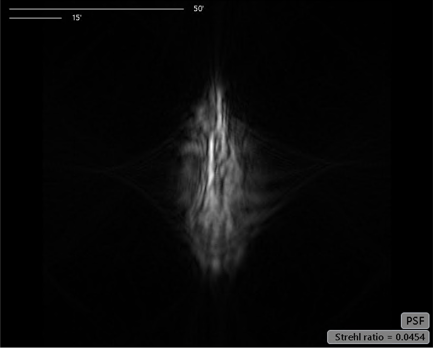

Astigmatism causes the PSF to become elongated. This causes ghosting and blurring of the vision. The same person with the astigmatism corrected.
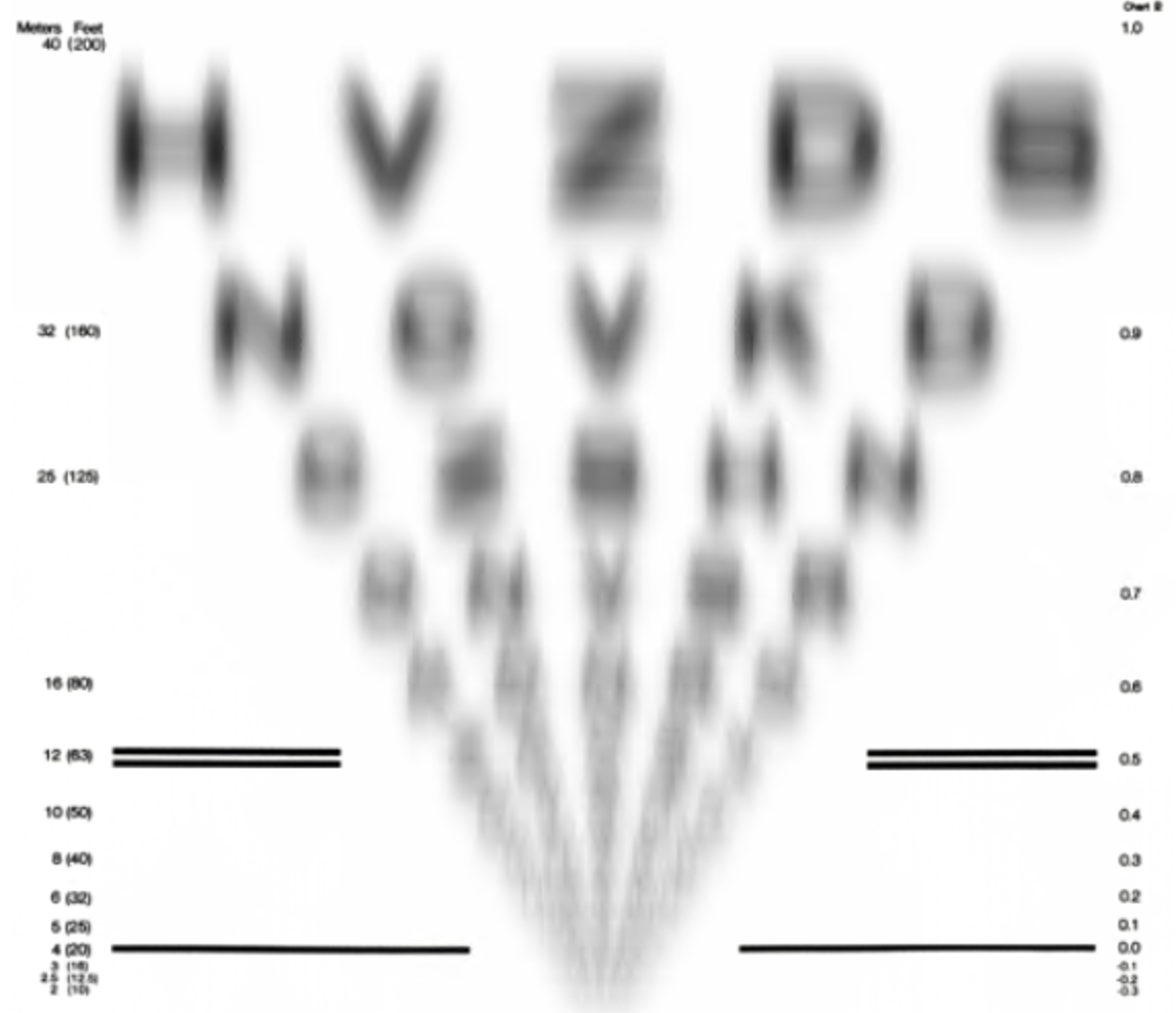

The vision chart becomes clear when the astigmatism is corrected.
Keratoconus
Keratoconus causes irregular astigmatism that is not fully correctable by glasses. The sliders below depict the before and after of the PSF and letter chart using glasses to correct the vision. Methods other than glasses must be used to fix the vision in keratoconus. See The repair process below.
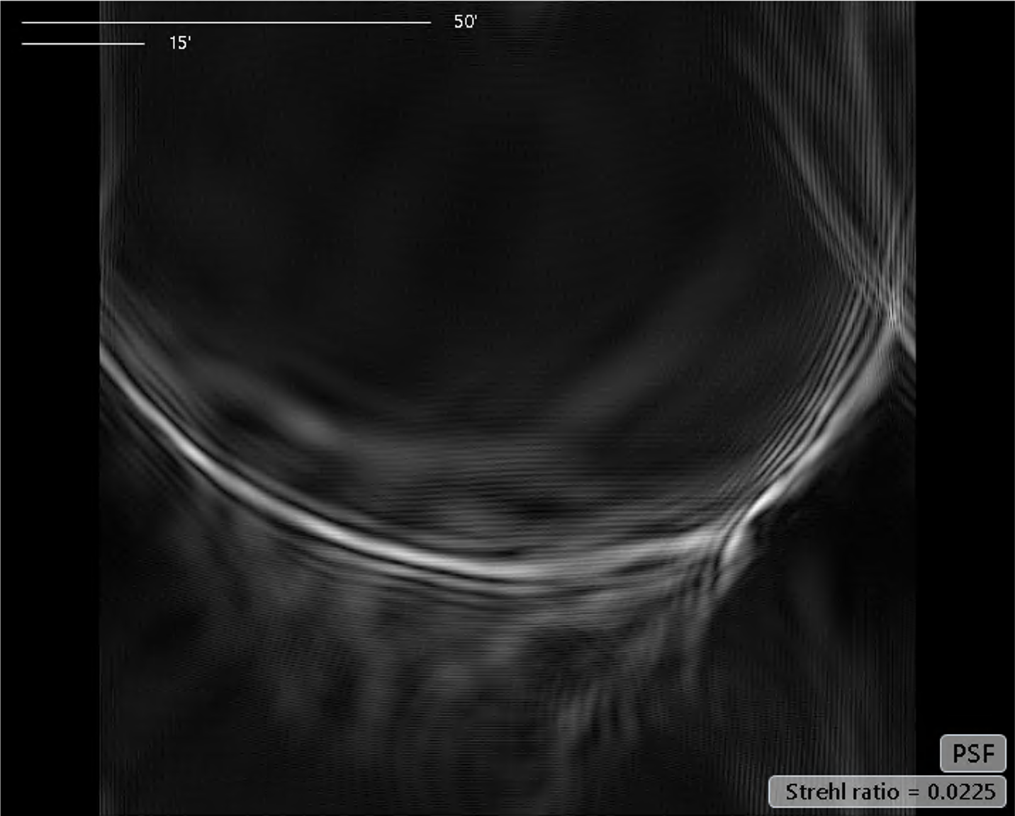
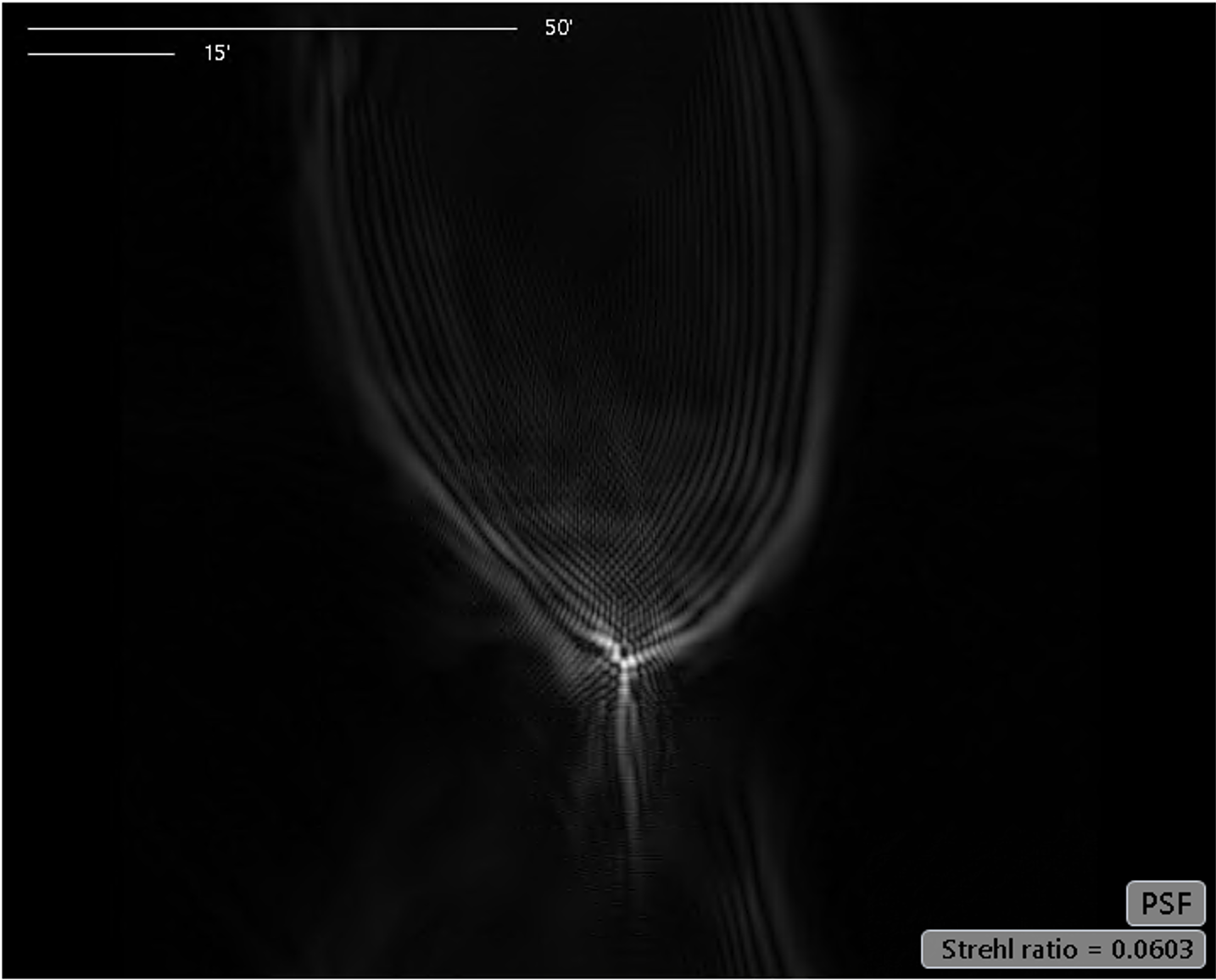
Observe how the PSF changes shape with an optimal glasses prescription, but it is not corrected. This glasses corrected PSF is a typical shape for keratoconus, resembling a comet and its dust trail in space.
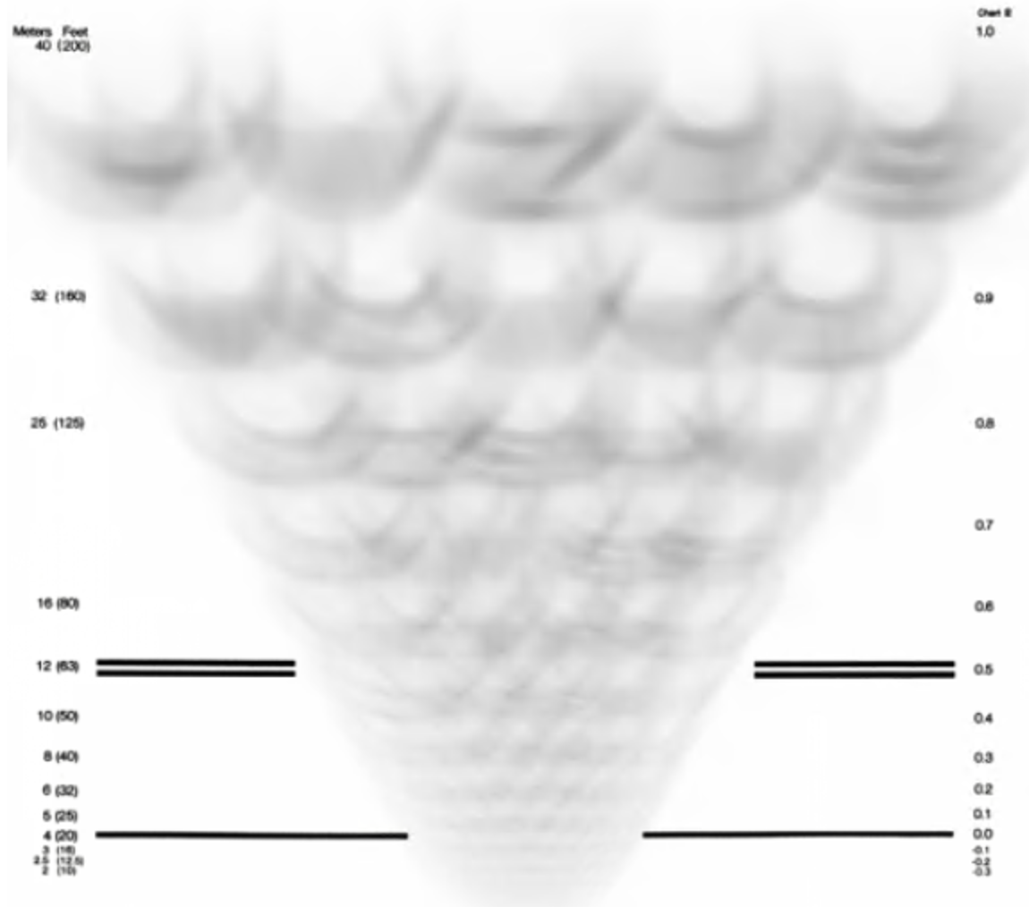
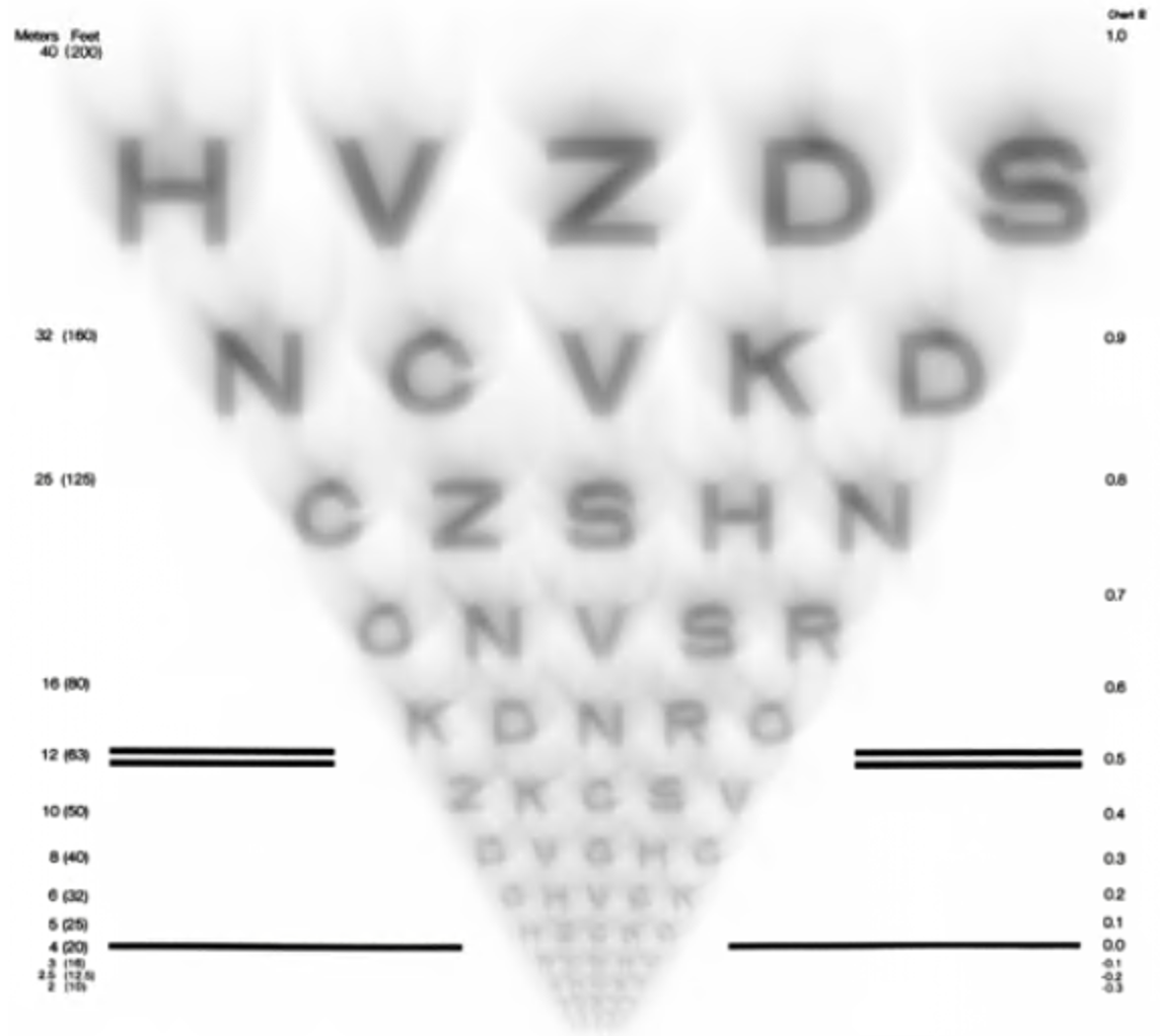
The slider shows how glasses improve the letter chart, but the characters remain slurred and blurred.
The repair process
During an initial assessment, detailed investigations and examinations are performed. Then, a treatment pathway is suggested. Treatment pathways typically have several stages, with a few months in between. The entire process may take several months to complete.
The first stage will depend on many factors, including the stage of your disease, the thickness of your cornea, and its optical clarity. Please use the link for further information on all of the options.
Customised laser eye surgery
Detailed scans are utilised to create a repair profile for the excimer laser for phototherapeutic keratectomy (PTK) — the excimer laser works by highly accurately removing tissue. The principles of the treatment are;
- To improve the shape of the cornea, ideally, creating one of the two regular shapes above.
- To minimise the amount of tissue removed
- To enhance the quality of vision
Surface laser eye surgery is used to apply the treatment to the cornea. This is a non-touch technique.
Treatment Timeline
The following timeline is for illustrative purposes only. A bespoke treatment pathway is generated for each eye.
Assessment
Initial assessment is completed and treatment plan formulated.
Managing vision
The repair process may make the vision worse before it is ultimately improved. We often use specially pre-fitted contact lenses for people who need to maintain vision throughout the process. This is especially the case if you choose to treat both eyes simultaneously to shorten the repair process.
Step 1 - First treatment
The first treatment is performed. If this is a customised laser or CAIRS, a minimum of three months is required for the healing process.
Step 2 - Potential second treatment
We may opt to perform a second treatment based on the outcomes of the first treatment. This is often the safest and most effective way for a more pronounced shape improvement.
Step 3 - Improve spectacle independence
The final stage is optional. Steps 1-2 aim to improve the quality of vision in glasses. Many patients prefer to reduce their dependence on glasses at this stage. Many options are available to achieve this, including, lens replacement surgery, implantable contact lenses, laser eye surgery.
Corneal scar
This is a real-life example of a patient treated by Mr Darcy for cornea scarring using this technique. The slider shows the before and after tomography shape scans, showing the efficacy of the treatment.

Previous decentred ablation
This individual had laser eye surgery in the 1990s abroad. He had suffered from poor vision since the procedure. He saw Mr Darcy, who instigated a multi-stage repair process. The slider shows the before and after scans, highlighting the first step of using customised laser eye surgery to 'undo' and repair the previous laser eye surgery. Following lens replacement surgery, this person now enjoys a new lease of life with restored vision.
Previous decentred ablation
This individual had laser eye surgery in the 1990s abroad. He had suffered from poor vision since the procedure. He saw Mr Darcy who instigated a multi-stage repair process. These scans highlight the first step of using customised laser eye surgery to 'undo' and repair the previous laser eye surgery. Following lens replacement surgery this person now enjoys a new lease of life with restored vision.
Keratoconus
This slider shows the before and after scans of a patient treated with Keratoconus. `the after scan shows regular astigmatism - a figure of eight shape. The prescription for astigmatism and residual glasses can then be managed conservatively (glasses or contact lenses) or with a permanent surgical solution to avoid needing contact lenses or glasses (Implantable contact lenses).

Keratoconus
This real-world example shows the before and after scans treating keratoconus. Removing tissue changes the glasses prescription. This patient preferred to be free of glasses and so following the customised laser had an implantable contact lens inserted.
Keratoconus
This real-world example slider shows the before and after scans treating keratoconus. Removing tissue changes the glasses prescription. This patient preferred to be free of glasses and so following the customised laser had an implantable contact lens inserted.
All eye surgical procedures carry a level of risk including not obtaining the desired outcome through to varying levels of visual loss. Your eye surgeon will discuss the risks, benefits and alternatives of sight correction surgery, including those specific to your own circumstances, at the time of your preoperative consultation